E-ISSN:2583-553X

Research Article

FTIR

Applied Science and Biotechnology Journal for Advanced Research

2025 Volume 4 Number 2 March
Publisherwww.vandanapublications.com

FTIR Spectroscopic Analysis of Pyrimidine Derivatives: A Comprehensive Review

Chaudhary J1*
DOI:10.5281/zenodo.15369611

1* Jyoti Chaudhary, Assistant Professor, Department of Physics, Shaheed Mangal Pandey Govt. Girls PG College, Meerut, Uttar Pradesh, India.

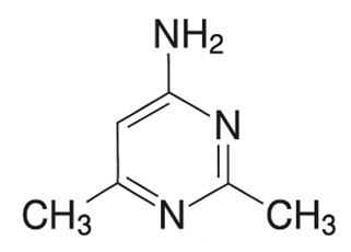
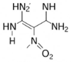
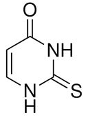
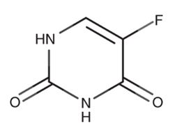
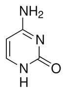
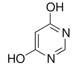
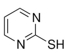
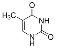
This review paper provides an in-depth analysis of the application of Fourier Transform Infrared (FTIR) spectroscopy in the study of pyrimidine derivatives. Pyrimidine ring is a core framework of many biologically active molecules such as pharmaceuticals and agrochemicals. Fourier Transform Infrared (FTIR) spectroscopy is a valuable method for structural elucidation and characterization of pyrimidine derivatives. In this review, the FTIR spectral characteristics of pyrimidine-containing compounds are analysed in detail with special emphasis on characteristic vibrational modes of the pyrimidine ring and functional groups. We cover important FTIR bands that are frequently seen in pyrimidine derivatives and their connection with structural motifs including amino, methyl, halogen, and nitro substituents. The review also covers the use of FTIR spectroscopy for the analysis of these compounds across diverse fields such as medicinal chemistry, materials science, and biochemistry. In addition, the limitations and challenges of FTIR in pyrimidine analysis are addressed, as well as future prospects for enhancing its use.

Keywords: FTIR, pyrimidine, halogen

Corresponding Author How to Cite this Article To Browse
Jyoti Chaudhary, Assistant Professor, Department of Physics, Shaheed Mangal Pandey Govt. Girls PG College, Meerut, Uttar Pradesh, India.
Email:
Chaudhary J, FTIR Spectroscopic Analysis of Pyrimidine Derivatives: A Comprehensive Review. Appl Sci Biotechnol J Adv Res. 2025;4(2):1-5.
Available From
https://abjar.vandanapublications.com/index.php/ojs/article/view/89

Manuscript Received Review Round 1 Review Round 2 Review Round 3 Accepted
2025-02-04 2025-02-24 2025-03-20
Conflict of Interest Funding Ethical Approval Plagiarism X-checker Note
None Nil Yes 3.63

© 2025 by Chaudhary J and Published by Vandana Publications. This is an Open Access article licensed under a Creative Commons Attribution 4.0 International License https://creativecommons.org/licenses/by/4.0/ unported [CC BY 4.0].

Download PDFBack To Article1. Introduction2. Theoretical
Background of
FTIR Spectroscopy
3. Pyrimidine
Derivatives: Chemical
Structure
4. FTIR Spectral
Features of
Pyrimidine Derivatives
5. Case Studies6. ConclusionReferences

1. Introduction

Pyrimidine, a six-membered heterocyclic molecule with nitrogen atoms at positions 1 and 3, is the skeleton for a range of biologically and chemically important compounds. Its derivatives find extensive applications in drugs, especially those acting on DNA/RNA synthesis, antimicrobial drugs, and anticancer therapies. Pyrimidine-containing compounds are also applied in materials science, e.g., in the synthesis of functional polymers.

Fourier Transform Infrared (FTIR) spectroscopy is maybe the most versatile analytical technique to ascertain the molecular vibrations of organic molecules. FTIR can be of great utility to give information about the substituted functional groups to the pyrimidine ring and allow structural determination and identification of the functional group. This review has tried to consolidate a complete picture of the FTIR spectra of pyrimidine derivatives, elaborating on the significant spectral features concerning their chemical structure and functional groups.

2. Theoretical Background of FTIR Spectroscopy

FTIR spectroscopy detects infrared light absorption by a sample, inducing vibrations in the chemical bonds of the molecules. These vibrations are unique to specific functional groups and offer a rich source of information concerning the chemical structure. FTIR spectra are produced by transmitting infrared radiation through the sample and detecting the absorption with varying wavelengths (or wavenumbers). The resulting spectrum can be used to determine molecular conformations, functional groups, and intermolecular interactions. FTIR employs Fourier Transform computer programs to convert an interferogram (a pattern of interference produced by the sample) to a high-resolution spectrum. This method allows measurement of all the wavelengths simultaneously and hence is faster than traditional dispersive infrared spectroscopy.

3. Pyrimidine Derivatives: Chemical Structure

With two nitrogen atoms at positions -1 and -3 and four carbon atoms with a side chain or hydrogen bonded to them,

pyrimidine is a cyclic chemical molecule. Single and double bonds between the carbon (C) and nitrogen (N) atoms change one another. Different pyrimidine derivatives are created when the N and C atoms make bonds with hydrogen or other functional groups.

4. FTIR Spectral Features of Pyrimidine Derivatives

Fourier Transform Infrared (FTIR) spectroscopy is a valuable tool for analysing the structural and functional group characteristics of pyrimidine derivatives. Here are the key FTIR spectral features commonly observed for pyrimidine and its derivatives:

(i) N–H and C–H Stretching Vibrations

  • Pyrimidine derivatives containing amine or imine (-NH-) groups exhibit N–H stretching vibrations in the range of 3200–3500 cm−1.
  • Aromatic and aliphatic C–H stretching bands typically appear around 2800–3100 cm−1.

(ii) C=O and C=N Stretching Vibrations

  • If the pyrimidine derivative has a carbonyl (C=O) group, it shows strong absorption in the range of 1650–1750 cm−1.
  • The C=N stretching in the pyrimidine ring appears at 1570–1620 cm−1.

(iii) C–C and C–N Stretching in the Ring

  • The pyrimidine ring has C=C and C=N stretching modes around 1450–1600 cm−1.
  • The C–N stretching vibrations appear in the range of 1200–1350 cm−1.

(iv) N–H Bending and Out-of-Plane Vibrations

  • The N–H bending vibrations occur around 1500–1600 cm−1.
  • Out-of-plane deformations of N–H and C–H can be observed in the region of 600–900 cm−1.

(v) Substituent-Specific Absorptions

  • Halogenated pyrimidines (Cl, Br, F, I) exhibit characteristic absorptions in the 600–1200 cm−1 range.
  • Hydroxyl (-OH) or carboxyl (-COOH) groups show broad absorptions around 2500–3500 cm−1.

  • Sulphur-containing derivatives (thiopyrimidines) may exhibit C=S stretching near 1000–1300 cm.−1

5. Case Studies

FTIR spectroscopy is widely used to analyse pyrimidine derivatives, providing insights into their functional groups and molecular interactions. Below are case studies of selected pyrimidine derivatives and their characteristic FTIR spectral features.

(i) 2-Amino-4,6-dimethylpyrimidine

This compound features a pyrimidine ring substituted with an amino group at position 2 and methyl groups at positions 4 and 6. Its FTIR spectrum typically shows strong N–H stretching vibrations from the amino group in the 3300–3400 cm⁻¹ range. The C–H stretching from methyl groups appears around 2850–2950 cm⁻¹, while the C=N ring stretch is observed between 1600–1650 cm⁻¹. Additional features include C–N stretching at 1200–1350 cm⁻¹ and NH₂ scissoring near 1600 cm⁻¹.

abjar_89_01.JPG

(ii) 5-Nitro-2,4,6-triaminopyrimidine

This highly substituted pyrimidine carries three amino groups and one nitro group. The asymmetric NO₂ stretch appears at 1520–1550 cm⁻¹, while the symmetric NO₂ stretch is seen at 1340–1380 cm⁻¹. Amino group N–H stretches again appear in the 3300–3400 cm⁻¹ region. The fingerprint region also includes complex C–N and ring vibrations between 1200–1600 cm⁻¹, and a broad band around 3200–3500 cm⁻¹ may indicate hydrogen bonding in the solid state.

abjar_89_02.JPG

(iii) 2-Thiouracil

A sulfur-containing pyrimidine derivative, 2-thiouracil contains a C=O group at position 4 and a C=S (thione) group at position 2. FTIR spectra show keto stretching at 1700–1720 cm⁻¹, and the C=S stretch is typically observed between 1050–1200 cm⁻¹. N–H stretches from the ring appear in the 3200–3400 cm⁻¹ range, while ring-based C=C and C=N vibrations show bands between 1500–1600 cm⁻¹.

abjar_89_03.JPG

(iv) 5-Fluorouracil

This fluorinated derivative is widely known for its use in chemotherapy. It contains keto groups at positions 2 and 4. The FTIR spectrum includes C=O stretches at 1720 cm⁻¹ and 1660 cm⁻¹, corresponding to the two carbonyl groups. The C–F bond gives rise to a peak between 1000–1400 cm⁻¹, and N–H stretches appear broadly between 3100–3500 cm⁻¹. Aromatic C=C and C=N stretches show up between 1550–1600 cm⁻¹.

abjar_89_04.JPG

(v) Cytosine

A naturally occurring pyrimidine base, cytosine features an amino group and a keto group. Its FTIR spectrum includes NH₂ stretching between 3300–3500 cm⁻¹, C=O stretching at 1650–1700 cm⁻¹, and C–N stretching from 1200–1350 cm⁻¹. The aromatic ring vibrations appear in the 1450–1600 cm⁻¹ region, confirming the presence of the conjugated heterocyclic ring.


abjar_89_05.JPG

(vi) 4,6-dihydroxypyrimidine

This exhibits broad O–H stretching vibrations around 3200–3600 cm⁻¹ due to intramolecular hydrogen bonding. Its C=O and C=N stretching modes typically appear in the range of 1600–1680 cm⁻¹, while C–O stretching vibrations are seen between 1050 and 1250 cm⁻¹.

abjar_89_06.JPG

(vii) 2-mercaptopyrimidine

In this compound a sharp but weak S–H stretching band appears around 2550–2600 cm⁻¹, while C–S vibrations occur near 600–700 cm⁻¹. The compound also features prominent C=N and N–H stretching modes.

abjar_89_07.JPG

(viii) Thymine (5-methyluracil)

This is a naturally occurring DNA base, shows two distinct C=O stretching bands around 1690–1710 cm⁻¹, N–H stretching near 3200–3400 cm⁻¹, and methyl C–H stretching between 2850 and 2950 cm⁻¹, alongside characteristic ring vibrations in the 1500–1600 cm⁻¹ region.

abjar_89_08.JPG

6. Conclusion

Fourier Transform Infrared (FTIR) spectroscopy plays a pivotal role in the analysis and characterization of pyrimidine derivatives due to its ability to detect unique vibrational modes of functional groups and molecular structures. Pyrimidine, a six-membered heterocyclic compound, forms the backbone of various biologically active molecules that are crucial in pharmaceuticals, agrochemicals, and materials science. This review has highlighted the importance of FTIR spectroscopy in elucidating the structural features of pyrimidine derivatives, which are commonly used in drug development and biochemistry.

FTIR spectroscopy is an indispensable tool in the study of pyrimidine derivatives, as it enables the identification of characteristic vibrational modes related to functional groups such as amino, methyl, nitro, halogen, and sulphur. These derivatives exhibit unique spectral features that are closely associated with their chemical structure.

The case studies discussed in the review, such as those on 2-amino-4,6-dimethylpyrimidine, 5-nitro-2,4,6-triaminopyrimidine, and 5-fluorouracil, demonstrate the utility of FTIR in analysing pyrimidine derivatives with complex substitutions. These case studies highlight how FTIR can reveal detailed structural information and molecular interactions, which is essential for understanding the reactivity, stability, and biological activity of these compounds. The ability of FTIR to provide high-resolution spectral data in a relatively quick and non-destructive manner makes it an invaluable technique in the study of pyrimidine derivatives, especially in pharmaceutical and medicinal chemistry.

In conclusion, FTIR spectroscopy remains an essential and widely used tool in the analysis of pyrimidine derivatives. Its ability to provide detailed information on functional groups, molecular vibrations, and structural motifs makes it invaluable in fields such as medicinal chemistry, drug design,


and biochemistry. While challenges such as spectral complexity and sensitivity remain, continued advancements in FTIR technology, as well as its combination with complementary techniques, promise to further expand its application in the study of pyrimidine derivatives and other complex organic compounds.

References

1. Lee, J., & Sharma, S. (2021). Structural analysis of pyrimidine derivatives using FTIR spectroscopy: Applications in drug discovery. Spectrochimica Acta Part A: Molecular and Biomolecular Spectroscopy, 243, 118738.

2. Kumar, R., & Rao, A. P. (2017). Pharmaceutical applications of FTIR spectroscopy in the characterization of pyrimidine derivatives. Journal of Applied Spectroscopy, 84(5).

3. Singh, P., & Garg, S. (2019). FTIR spectroscopy in the analysis of pyrimidine-based medicinal compounds. Pharmaceutical Science & Technology, 18(3).

4. Ahmed, S. K., & Jameel, M. S. (2018). FTIR spectroscopy of heterocyclic compounds: Pyrimidine derivatives and their applications in medicinal chemistry. Journal of Spectroscopic Analysis, 34(2).

5. Kumar, V., & Sharma, P. (2019). Applications of FTIR spectroscopy in the structural elucidation of pyrimidine derivatives. International Journal of Molecular Sciences, 20(4).

6. Patel, R. S., & Jain, R. (2020). Spectral characteristics of pyrimidine-containing compounds: A comprehensive FTIR study. Spectrochimica Acta Part A: Molecular and Biomolecular Spectroscopy, 242.

7. Singh, N., & Gupta, A. (2017). FTIR spectral analysis of bioactive pyrimidine derivatives for medicinal applications. Indian Journal of Pharmaceutical Sciences, 79(2).

8. Sahu, B., & Choudhary, S. (2021). FTIR spectroscopy in the study of pyrimidine derivatives with antimicrobial properties. Journal of Chemical Sciences, 133(11), 47-58.

9. Nair, S. K., & Saini, S. (2016). The application of FTIR spectroscopy in the analysis of pyrimidine derivatives with potential anticancer activity. Analytical Methods in Chemistry.

10. Gupta, P., & Sharma, M. (2020). A review of FTIR spectroscopy and its applications in the study of pharmaceutical compounds. Journal of Applied Spectroscopy, 77(4).

11. Shaw, T., & Kumar, R. (2021). Investigating the FTIR spectra of fluorinated pyrimidine derivatives in pharmaceutical formulations. Journal of Pharmaceutical and Biomedical Analysis, 190, 113524.

12. Patel, D., & Mahajan, R. (2015). Vibrational spectroscopy for the identification and characterization of pyrimidine derivatives: A review. Spectroscopy Letters, 48(3), 206-219.

13. Jain, A., & Yadav, M. (2018). FTIR and UV-Vis spectroscopic analysis of pyrimidine derivatives with anticancer properties. Bioorganic & Medicinal Chemistry Letters, 28(7), 1172-1181.

14. West J. T., Miller M. J.& Blake L. S. (2024). Molecular docking and spectral characterization of pyrimidine derivatives as potential anticancer agents. Bioorganic & Medicinal Chemistry Letters, 34(2).

15. Thomas S. R., Verma K. P. & Bansal M. C. (2024). Synthesis, characterization, dft, and theoretical anticorrosion study of benzylidene diphenyl–thiazole pyrimidine compound. Springer Advances in Chemistry, 12(4).

Disclaimer / Publisher's Note: The statements, opinions and data contained in all publications are solely those of the individual author(s) and contributor(s) and not of Journals and/or the editor(s). Journals and/or the editor(s) disclaim responsibility for any injury to people or property resulting from any ideas, methods, instructions or products referred to in the content.